Dynamics,
structure and
molecular biology
of fungal invasion

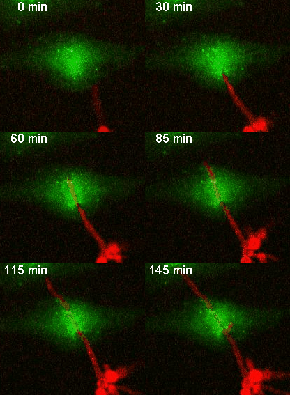
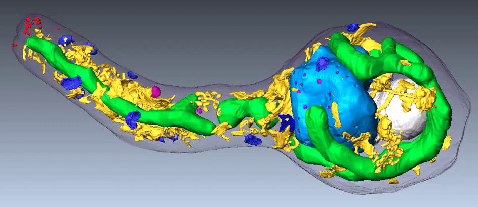
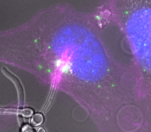
Our ATIP-Avenir team, established in 2018, studies the invasion mechanisms of the human fungal pathogen Candida albicans in epithelial layers using live cell imaging, cell biology and correlative light and electron microscopy.
By integrating dynamics, structure, and cellular and molecular biology, we address fundamental questions regarding how fungal infections are initiated and lead to disease.
The team members
Our research focus on 3 axes
- Pathogenic fungi
- Invasion of Candida albicans
- Live cell imaging
- Optical and correlative electron microscopy
- Cell biology
- Cellular microbiology
1. Fungal invasion of epithelial tissue
Fungal pathogens are responsible for as many deaths each year as tuberculosis or malaria. The opportunistic fungal pathogen Candida albicans colonises the skin, genital and intestinal mucosa of most healthy individuals and is part of the normal commensal flora. In susceptible hosts, C. albicans can invade the gastrointestinal mucosa and enter the bloodstream, resulting in severe systemic infection.
2. Interactions between the pathogen and the host
Invasion by C. albicans is a challenging area of investigation, as it encompasses a complex set of interspecies interactions shaped over millions of years of evolution in an ‘arms race’. Events at this interface are often highly transient and involve the pathogen’s subversion of host pathways and the activation of host defence mechanisms. Understanding the invasion of C. albicans is a major challenge, requiring a combination of dynamic, structural and molecular approaches.
3. Integrating dynamics, structure and molecular information to study infection
The aim of our laboratory is to study the invasion mechanisms of Candida albicans in epithelial layers at the single cell level using multidimensional fluorescent microscopy, cell biology and correlative light and electron microscopy.
The opportunities
- Mechanisms of infection by Candida albicans
- Understanding the host and pathogen molecular factors that regulate fungal infection
Publications
Trans-cellular tunnels induced by the fungal pathogen Candida albicans facilitate invasion through successive epithelial cells without host damage. Nat Commun. 2022 Jun 30;13(1):3781. doi: 10.1038/s41467-022-31237-z. Free PMC article.
Step by step guide to post-acquisition correlation of confocal and FIB/SEM volumes using Amira software. Weiner A. Methods in cell biology: Correlative Light and Electron Microscopy (2020) IV Volume 1622020.
Fundamental Helical Geometry Consolidates the Plant Photosynthetic Membrane. Bussi Y., Shimoni E., Weiner A., Kapon R., Charuvi D., Nevo R., Efrati E., Reich Z.
Proc Natl Acad Sci U S A. 2019
The pathogen-host interface in three-dimensions: correlative FIB/SEM applications. Weiner A. and Enninga J.Trends Microbiol. 2019
On-site secretory vesicle delivery drives filamentous growth in the fungal pathogen Candida albicans. Weiner A., Orange F., Lacas-Gervais S., Rechav K., Ghugtyal V., Bassilana M., Arkowitz R.A. Cell Microbiol., 2019


